您的位置: 主页 > 产品展示 > 细胞成像工具 >

ACC-103 Anti-CACNA2D3 (CaVα2δ3) (extracellular) Antibody

发布日期:2021-01-19 14:34   浏览量:
Cat #: ACC-103
Host: Rabbit
Type: Polyclonal
Form: Lyophilized

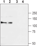
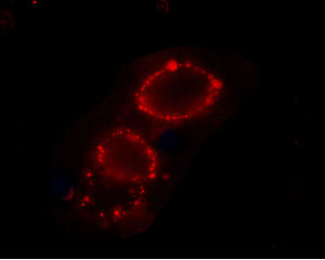
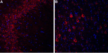
Anti-CACNA2D3 (CaVα2δ3) (extracellular) Antibody (#ACC-103) is a highly specific antibody directed against an extracellular epitope of the rat protein. The antibody can be used in western blot, immunohistochemistry and immunocytochemistry applications. It has been designed to recognize CaVα2δ3 from rat, mouse and human samples.


Applications: ICC, IF, IHC, LCI, WB
May also work in: IFC, IP
Reactivity: H, M, R

Application key:

 CBE- Cell-based ELISA, FC- Flow cytometry, ICC- Immunocytochemistry, IE- Indirect ELISA, IF- Immunofluorescence, IFC- Indirect flow cytometry, IHC- Immunohistochemistry, IP- Immunoprecipitation, LCI- Live cell imaging, N- Neutralization, WB- Western blot

Species reactivity key:

 H- Human, M- Mouse, R- Rat
 
 
For research purposes only, not for human use

Applications

 
Western blot
  • Western blot analysis of rat brain (lanes 1 and 3) and K562 (lanes 2 and 4) lysates:
    Western blot analysis of rat brain (lanes 1 and 3) and K562 (lanes 2 and 4) lysates:
    1,2. Anti-CACNA2D3 (CaVα2δ3) (extracellular) Antibody (#ACC-103), (1:200).
    3,4. Anti-CACNA2D3 (CaVα2δ3) (extracellular) Antibody, preincubated with CACNA2D3/Cavα2δ3 (extracellular) Blocking Peptide (#BLP-CC103).
Live cell imaging / Immunocytochemistry
  • Expression of CaVα2δ3 in rat PC12 cells
    Expression of CaVα2δ3 in rat PC12 cells
    Cell surface detection of CaVα2δ3 in intact living PC12 cells. Extracellular staining of cells using Anti-CACNA2D3 (CaVα2δ3) (extracellular) Antibody (#ACC-103), (1:25) followed by goat anti-rabbit-AlexaFluor-594 secondary antibody.
Immunohistochemistry
  • Expression of CaVα2δ3 in rat hippocampus and cortex
    Expression of CaVα2δ3 in rat hippocampus and cortex
    Immunohistochemical staining of rat hippocampal CA3 region (A) and rat neocortex (B) using Anti-CACNA2D3 (CaVα2δ3) (extracellular) Antibody (#ACC-103). In both areas CaVα2δ3 staining (red) appears in pyramidal neurons (arrow). DAPI is used as the counterstain (blue).

在线咨询 联系方式 二维码

服务热线

021-50724187

扫一扫,关注我们