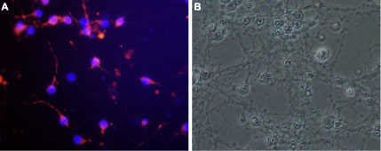
Expression of Nogo receptor in rat cerebellar granule
Cell surface detection of Nogo receptor in live cultured rat cerebellar granule. A. Cells were stained with Anti-Nogo Receptor (extracellular) Antibody (#ANT-008) followed by goat-anti-rabbit AlexaFluor-555 secondary antibody (red). Nuclei were visualized with the cell permeable dye Hoechst 33342 (blue). B. Live view of the same field as in A.
Immunohistochemistry
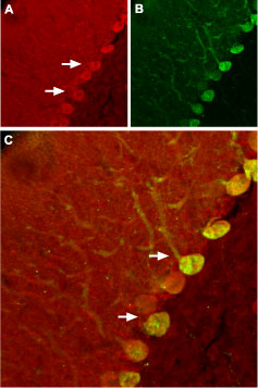
Expression of Nogo receptor in rat cerebellum
Immunohistochemical staining of rat cerebellum using Anti-Nogo Receptor (extracellular) Antibody (#ANT-008). A. NgR (red) appears in Purkinje cells (arrows). B. Staining of Purkinje nerve cells with mouse anti-calbindin D28K (a calcium binding protein, green). C. Confocal merge of NgR and calbindin D28K demonstrates some co-localization of these proteins.
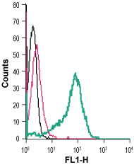
Indirect flow cytometry
Cell surface detection of Nogo Receptor in live intact human THP-1 monocytic leukemia cells: