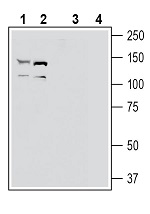
Western blot analysis of human U87-MG glioblastoma cell line lysate (lanes 1 and 5), human THP-1 monocytic leukemia cell line lysate (lanes 2 and 6), human LNCaP prostate adenocarcinoma cell line lysate (lanes 3 and 7) and human MCF-7 breast adenocarcinoma cell line lysate (lanes 4 and 8):
Immunohistochemical staining of perfusion-fixed frozen rat brain sections with Anti-EphA2 (extracellular) Antibody (#AER-019), (1:300), followed by goat anti-rabbit-AlexaFluor-488. A. Staining in the rat hippocampal CA1 region, showed immunoreactivity (green) in neuronal profiles in both soma (vertical arrows) and apical dendrites of pyramidal neurons (horizontal arrows). B. Pre-incubation of the antibody with EphA2 (extracellular) Blocking Peptide (#BLP-ER019), suppressed staining. Cell nuclei are stained with DAPI (blue). P = pyramidal layer, SO = stratum oriens, SR = stratum radiatum.
Expression of EphA2 in mouse cortex.
Immunohistochemical staining of perfusion-fixed frozen mouse brain sections with Anti-EphA2 (extracellular) Antibody (#AER-019), (1:300), followed by goat anti-rabbit-AlexaFluor-488. A. EphA2 immunoreactivity (green) appeared in neuronal profiles in both soma (vertical arrows) and apical dendrites of pyramidal neurons (horizontal arrows). B. Pre-incubation of the antibody with EphA2 (extracellular) Blocking Peptide (#BLP-ER019), suppressed staining. Cell nuclei are stained with DAPI (blue).
Indirect flow cytometry
Cell surface detection of EphA2 in live intact human THP-1 monocytic leukemia cells: